Abstract
Introduction:
DYT-11 is a form of myoclonus dystonia (MD) characterized by involuntary muscle jerks and abnormal postures attributable to a variant in the epsilon sarcoglycan (SGCE) gene. Treatment with pallidal deep brain stimulation (GPi-DBS) is effective, but prior studies have highlighted brisk and facile responses to stimulation. While medically refractory cases are common, the literature lacks cases refractory to initial surgical therapy and there are no reports of advanced programming or DBS revision surgery. Our series aims to provide insight into the advanced management of these patients.
Methods:
Patients treated for genetically confirmed DYT-11 with DBS were identified. Retrospective chart review was performed.
Results:
We report two cases of DYT-11 sub-optimally responsive to DBS that were successfully treated with DBS revision surgery. Lead revision and subsequent programming provided a significant improvement in symptoms. We also report a case of a patient with DYT-11 who was successfully treated with DBS but required advanced programming to achieve best benefit.
Discussion:
We present three cases of DYT-11 that required advanced care to achieve successful treatment with DBS. These approaches have not previously been published in DYT-11 and highlight heterogeneity of response in this disorder. Further studies are needed to investigate optimal strategies for DBS troubleshooting in DYT-11 such as characterizing electrophysiology and brain connectomics.
Introduction
Myoclonus dystonia (MD) is characterized by involuntary muscle jerks and abnormal postures. MD caused by variants in the epsilon-sarcoglycan (SGCE) gene is autosomal dominant with incomplete penetrance and termed DYT-11 [1]. Classically, this condition produces a combination of myoclonus and dystonia with frequent psychiatric manifestations and alcohol-responsiveness, but the phenotype has some variability that extends to tremor and even stuttering [2, 3]. Deep brain stimulation (DBS) is a well-established therapeutic option for medically intractable MD [2, 3]. A recent review of DBS in MD found that 91.8% of patients had improvements in the motor Burke Fahn Marsden Dystonia Rating Scale (BFMDRS) and 79.6% had improvements of >50%. In most patients, therapeutic effect is achieved quickly with a monopolar stimulation configuration [3–6]. Overall, the literature suggests that MD is consistently briskly responsive to DBS. As a result, for patients with refractory symptoms, there is little guidance.
For dystonia at large, achieving control of symptoms with DBS can prove challenging. While there is no doubt this therapy is effective, achieving a good outcome often takes time and experimentation. There are many reasons for this, but the variability in reported effective parameters in the literature, lead locations, duration of stimulation required to elicit a response, and differences in side effect tolerances all likely contribute. Attempts to find pooled data to determine effective strategies have resulted in heterogeneity. There is some signal for differences between subtypes, such as a predisposition towards higher pulse widths in DYT-1 dystonia, but data is difficult to parse. To date, only basic settings have commonly been reported for myoclonus dystonia [7].
Methods
Patients treated with DBS for genetically confirmed DYT-11 at our center were identified. Retrospective chart review was performed.
All patients were implanted with Medtronic Percept PC implantable pulse generators (IPGs) allowing local field potential (LFP) recordings to be performed during routine DBS programming visits. LFP data were extracted as .JSON files and analyzed using the BRAVO platform using previously described methodology [8].
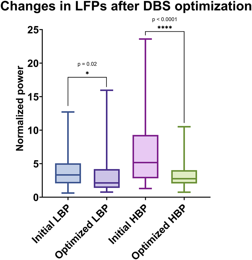
LFPs were recorded for 30 s at two different time points while patients were at rest. Spectral data was extracted from the raw time series using Welch’s periodogram, with a 2000 ms window and a 500 ms overlap, achieving a frequency resolution of 0.5 Hz. The spectral features were then normalized using the fitting oscillations and one over f (FOOOF) method. We defined the spectral features using the canonical definitions: low beta from 12 Hz to 20 Hz, high beta from 20 Hz to 30 Hz [9]. We employed a non-parametric Mann-Whitney U test to compare the initial and optimized normalized low beta power (LBP) and high beta power (HBP) spectral features across the three patients.
High resolution pre-operative MRI brain and post-operative CT head were obtained for each subject in this cohort. The Lead-DBS software package was used for image processing utilizing previously published methods [10]. Briefly, the post-operative high-resolution non-contrast brain CT was co-registered to the preoperative T1-weighted MPRAGE MRI brain using a two-stage linear registration using Advanced Normalization Tools (ANTs) [11]. The pre- and post-operative images were spatially normalized into MNI_ICBM_2009b_NLIN_ASYM template space using the symmetric normalization (SyN) registration approach. The DBS leads were spatially localized using the PaCER method within the Lead-DBS software, and manual verification and correction were applied if necessary [12].
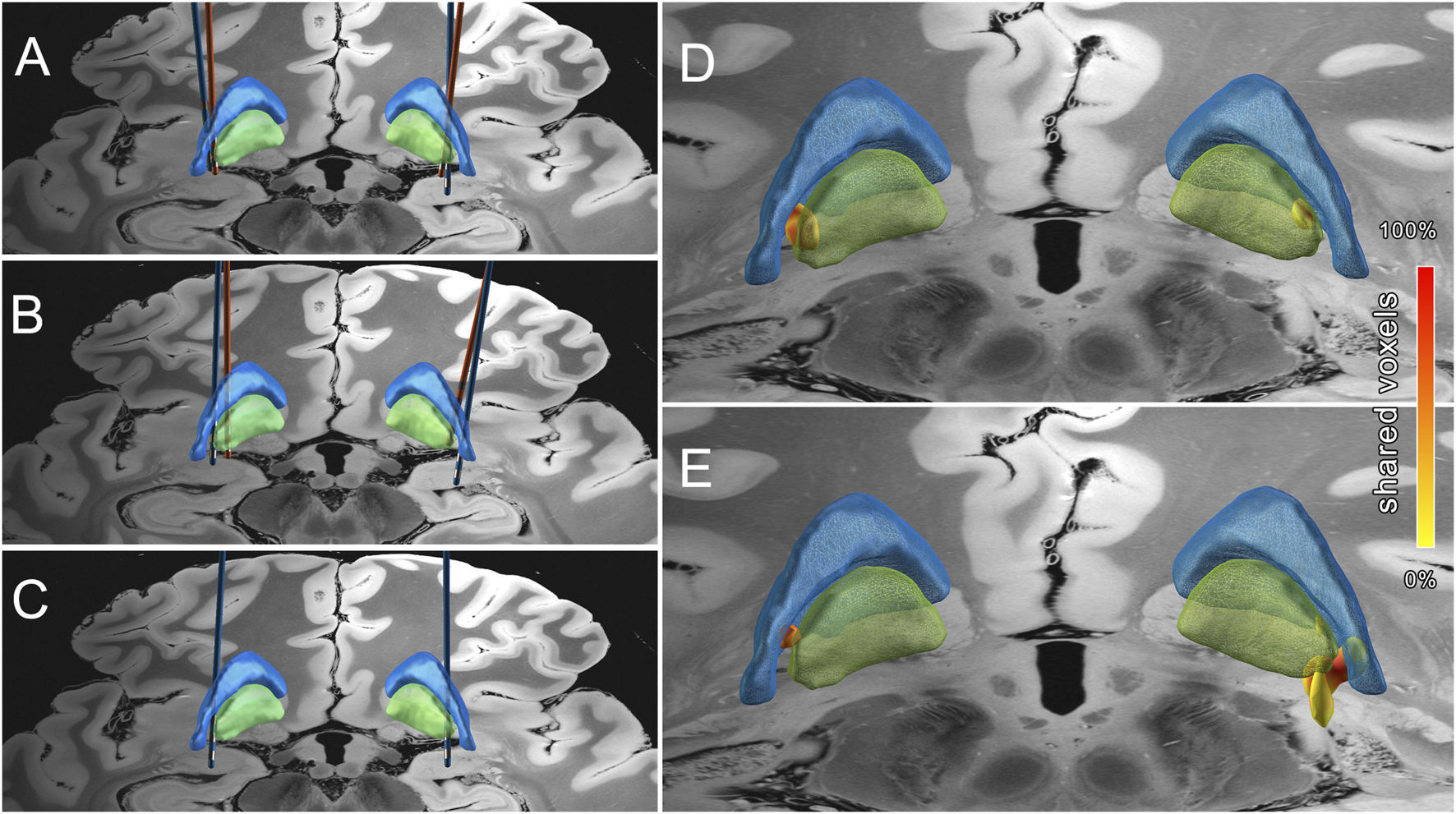
The volume of tissue activated (VTA) was estimated using finite-element modeling within the Lead-DBS software [10]. A binary VTA was generated over a tetrahedral mesh head model defined as an isotropic volume with a symmetric conductivity of 0.14 S/m and an electric field threshold of 0.2 V/mm [13]. A group level VTA heat map was created by summing the VTA voxels together using FSL’s fslmaths function to determine the most shared regions of stimulation (Figures 1D, E).
FIGURE 1

Lead locations before (orange) and after revision (blue) for MD01 (A), MD02 (B) and MD03 (C). The volume of tissue activated (VTA) is displayed based on pre-revision (D) and optimized settings (E). Overlapping voxels are represented by the color gradient with red representing total overlap and yellow no overlap. The globus pallidus internus is projected in green and the globus pallidus externus is projected in blue. Lead localization completed using Lead-DBS software suite [14].
Results
Case 1
MD01 is a 27-year-old woman with a family history of DYT-11 and a genetic test for a SGCE variant (c.835_839del) from birth who developed generalized myoclonus and cervical dystonia in early childhood. She underwent bilateral GPi-DBS (Medtronic 3389 leads) at age 18, but eventually experienced suboptimal symptom control. She was referred to our center for DBS troubleshooting. Her primary complaints were near-constant jerks and dystonic pain. MRI-based lead localization showed that the original leads were implanted reasonably in the posteromedial GPi, but resulted in low therapeutic windows and capsular side effects during monopolar review.
During advanced programming over 13 months, titration of stimulation amplitudes, pulse widths, and frequencies across a large range as well as bipolar contact configurations failed to provide satisfactory symptom control for both myoclonus and dystonia.
The patient and DBS team decided to proceed with bilateral GPi-DBS revision surgery (Medtronic B33015 leads). Three months post-revision, after a single post-operative programming session, dystonia-related pain and tension were completely resolved and myoclonus was significantly improved (resolution of stimulation-induced myoclonus and significant reduction in spontaneous myoclonus that worsened immediately with pausing therapy). The new lead locations were lateral and ventral to the prior lead locations, allowing tolerance of higher programming settings (Figure 1A). Dystonia severity measured by the BFMDRS in the DBS ON state improved from 21 pre-revision to 1 at 7 months post-revision. At 13 months post-revision BFMDRS was 3. DBS settings are summarized in Table 1.
TABLE 1
| Patient | Context | Left GPi | Right GPi |
|---|---|---|---|
| MD01 | Best settings prior to revision | 1- 2 + 2.5 mA 60 µs 210 Hz | 9–10 + 2.5 mA 60 µs 210 Hz |
| Best settings following revision | 1- C + 3.0 mA 140 µs 125 Hz | 9- C+ 1.7 mA 140 µs 125 Hz | |
| MD02 | Best settings prior to revision | Interleaving 1b- C + 3.5 V 60 µs 125 Hz 1–2 + 1.55 V 90 µs 125 Hz |
Interleaving 10–9 + 11+ 2.85 V 70 µs 125 Hz 8–11 + 0.6 V 60 µs 125 Hz |
| Best setting following revision | 2- C + 2.5 mA 90 µs 125 Hz | 10- C+ 1.5 mA 90 µs 125 Hz | |
| MD03 | Initial settings | 2- C + 1.5 mA 90 µs 135 Hz | 10- C+ 1.5 mA 90 µs 135 Hz |
| Final settings | 1- 2- C+ 2.3 mA 90 µs 165 Hz | 9–10- 8 + 3.9 mA 90 µs 165 Hz |
Summary of key DBS settings.
Case 2
MD02 is a 30-year-old woman who developed shaking of the head and hands at age 3. She was diagnosed with generalized dystonia at age 5 and genetic testing at age 23 revealed a variant in the SGCE gene (c.826-2A>G). Despite medical management, she became dependent for feeding and ambulation. At age 26, she underwent bilateral GPi-DBS (Medtronic 3387 leads). She improved rapidly, but later precipitously worsened and her device was explanted for concern for hardware infection. Bilateral GPi-DBS leads (Medtronic B33015) were reimplanted, but benefit was never fully recapitulated. She was referred to our center for DBS troubleshooting.
At our institution, monopolar review of her reimplanted leads revealed low thresholds for side effects. Notably, even at time of initial evaluation there was no significant burden of myoclonus. She presented with complex bipolar settings with interleaving with no perceived benefit (Table 1). Lead localization of her reimplanted leads revealed suboptimal lead location that was dorsal on the right and anteromedial on the left relative to the initial lead locations (Figure 1B). The combination of benefit with initial DBS leads, restrictive thresholds, and poor benefit despite complex programming motivated the decision to pursue DBS lead revision. While conducting a multidisciplinary pre-surgical evaluation, reprogramming with bipolar and multiple monopolar configurations yielded some improvement to neck dystonia, but significant debility remained with a United Dystonia Rating Scale (UDRS) score of 25.
She underwent repeat bilateral GPi-DBS revision (Medtronic B33015 leads) (Figure 1B). Post-revision, she had larger side effect thresholds and was programmed on simple monopolar settings (Table 1). One year post-revision there was no residual dystonia and mild residual right hand postural and kinetic tremor. At 23 months post-revision, benefit was sustained with UDRS of 13.
Case 3
Patient MD03 is a 75-year-old female who exhibited arm tremors in infancy followed by an insidious progression of dystonia and myoclonus spanning several decades that culminated in a late-life diagnosis of MD corroborated by positive genetic testing for a variant in the SGCE gene (c.551T>C). Multiple pharmacological interventions were limited by side effects. The patient sought advanced care at our institution.
The patient chose to undergo DBS implantation. Preoperative assessments revealed a Toronto Western Spasmodic Torticollis Rating Scale (TWSTRS) score of 38. Staged bilateral GPi-DBS surgery was performed (Medtronic 3387 leads). There was a stimulation-induced worsening of myoclonus in the operating room at the ventral-most contact during intraoperative physiology testing. Three months after the first lead implantation, the patient continued to have problematic dystonia and myoclonus was mildly improved. Initially, simple settings were used (Table 1). Pulse width modulation was ineffective. While increasing the amplitude produced some benefit, it also led to capsular side effects. Double monopolar settings were explored which did improve myoclonus, followed by additional amplitude adjustments made possible by leveraging a three-contact bipolar setting. Significant symptomatic benefit was achieved with complex settings (Table 1). Six months after the placement of the second GPi lead, her condition was significantly improved with a TWSTRS score of 14. After 12 months of stimulation, her TWSTRS score was 7, and at 33 months she reported no residual disability.
Local field potential analysis
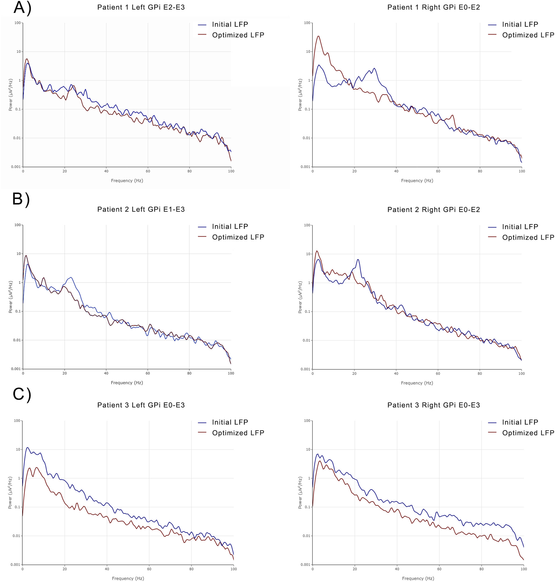
In MD01, a broad band beta peak was observed in both GPi regions following bilateral revision surgery. After optimizing the DBS settings, there was a notable reduction in the broad band beta signal. This reduction was more pronounced in the right GPi compared to the left GPi (Figure 2A).
FIGURE 2

PSDs from the left GPi before and after optimization and right GPi pre- and post-revision of MD01 (A), left and right GPi of MD02 pre- and post-revision (B), and left and right GPi of MD03 before and after optimization (C).
In MD02, both the left and right GPi LFPs demonstrated marked power in the beta band prior to DBS revision. Following revision and effective stimulation, there was a decrease in beta power (Figure 2B).
In MD03, both the right and left GPi LFPs initially demonstrated peaks in the theta/alpha and beta bands. Following DBS optimization, broadband power decreased bilaterally (Figure 2C).
These observations were confirmed using the previously described signal processing and statistical methods. There was a significant decrease in both HBP (p < 0.001) and LBP (p = 0.02) between the initial and post-optimization states (Figure 3).
FIGURE 3

Mean normalized low beta power (LBP) defined as 12–20 Hz activity in the initial state (blue) and optimized state (purple) and normalized high beta power (HBP) defined as 20–30 Hz activity in the initial state (pink) and optimized state (green) are displayed. The upper and lower whiskers of the boxplot represent 1.5 times the interquartile range.
Discussion
This is the first report of advanced programming and DBS revision in GPi-DBS for DYT-11. Prior studies of DBS in DYT-11 report dramatic improvement by the first recorded follow up and cite the need for only minimal adjustments to programming [6, 15–17]. A meta-analysis of DBS outcomes in MD reported significant improvement in dystonia at 0–6 months, but notably the median number of patients per study was 1 highlighting the rarity of this disease and potential susceptibility to publication bias [2].
In generalized dystonia at large, DBS takes time to produce maximal benefit and multiple programming sessions over months are often required [18]. This may be true of DYT-11 as well, particularly in cases with initial difficulty in achieving benefit.
Our approach is to begin with a monopolar review and if the thresholds for persistent side effects are uniformly less than 3 mA at a pulse width of 90 µs at a frequency of 130 Hz, we suspect difficultly in delivering sufficient energy to obtain benefit in dystonia.
Our first step in seeking benefit is to titrate current at this pulse width and frequency. Thereafter, we typically employ an increased pulse width, with care to avoid capsular side effects, followed by titration of frequency. Occasionally, lowering frequency can facilitate tolerance of higher pulse widths, but failing this we titrate up on frequency. Next, we employ bipolar and increasingly complex configurations including directional stimulation. Rarely, we will next attempt interleaving or cycling strategies. In general, we find interleaving and cycling more effective for patients who initially see signal for benefit and then quickly experience loss of benefit and find this less useful in cases of dystonia than in other etiologies treated with DBS.
A challenge in MD is that different phenomenology responds differently both in terms of time to benefit, where we expect a longer time to benefit for dystonia than myoclonus, and in terms of effect size, where classically the effect on myoclonus is more complete than that for dystonia. In our refractory cohort, myoclonus was more responsive to stimulation than dystonia. A potential pitfall for clinicians is to accept sub-optimal improvement in dystonia because of adequate control of myoclonus with a particular DBS programming configuration. Our cases highlight the real world situation wherein the first setting is often not the best setting in contrast to the current literature on DBS in MD.
If these approaches are unsuccessful, we consider lead revision or rescue leads. Lead location and the patient’s quality of life are core components in decision making. A key element is to ensure that the patient and care team align on expectations and goals for surgery.
MD is a complex disease with wide phenotypic variability. While larger amplitude jerks and myoclonus are more effectively managed with DBS, other symptoms such as dystonia, pain, kinetic tremor, and even retropulsion (as noted in one case report) may persist [16]. It is important to include this information during counseling. Informing patients that achieving benefits for dystonia may take a relatively longer time can help improve their satisfaction with the surgical outcome.
With the enhanced capabilities of modern DBS devices, chronic recording of electrophysiologic signals is now possible in the outpatient setting. This can serve as a tool to optimize DBS in challenging cases. Prior intraoperative data has shown correlation between low frequency oscillations (LFO) in the 3–15 Hz range in the GPi and involuntary EMG activity in the operating room [19, 20]. In other forms of dystonia there has been a similar correlation with manifest dystonia [21, 22]. More recently, a single case of DYT-1 dystonia reported benefit with programming to suppression of beta range oscillatory activity, further highlighting the importance of neurophysiologic signals as potential feedback mechanism to guide DBS programmers in dystonia [23]. However, for MD there remains a paucity of electrophysiology data from outside of the operating room. A single publication on artifact reduction included three MD patients. However, one patient was studied intraoperatively during battery replacement, another had contamination of the LFO band with tremor signals, and none had reported confirmatory genetic information [24]. In our patients, there were marked changes in LFPs after effective stimulation (Figure 2) that demonstrated a statistically significant decrease in both HBP and LBP at the group level (Figure 3). While this could have implications for programming, these results should be interpreted with caution as they represent an n of 3 and require confirmation through an analysis of a larger cohort. To our knowledge, we are the first to report these observations.
The clinical relevance of beta oscillatory activity in normal human physiology, in the more well described population of Parkinson’s disease, and in dystonia, remains debated. Posited roles in normal physiology, based on animal models and manipulation with drugs and electrical stimulation, have suggested a role for beta burst activity in integration of sensory information [25]. In the cortex there are associations with increased beta activity and movement cancellation and it has been suggested that beta activity can be conceptualized as a top-down inhibitory signal that enforces the “status quo” [25]. The suspected pathophysiology of dystonia at large has been hypothesized to relate to impaired sensorimotor integration and network level dysfunction and one can posit how aberrations in these signals could contribute [26]. However, there is very limited study of these factors in MD specifically and of beta signals in the pallidum as observed in our work, so whether these signals have clinical meaning requires further study. Further, some suggest that beta physiology is not increased among a heterogeneous population of dystonia patients, at least compared to those with Parkinson’s disease [27, 28]. In one study, dystonia patients were used as the control to highlight beta burst dynamics in Parkinson’s disease patients, due to the belief that patients with dystonia would not have elevated beta band power. In this instance, dystonia patients were likely selected because DBS provides access to pallidal signals and this small study did not include genetic dystonia [28].
Here, we demonstrate that beta band oscillatory activity may have a previously underrecognized role in DYT-11, warranting further exploration. However, the reasons this may be true in genetic dystonia, as recently shown in DYT-1, raise many questions. Further studies are needed to investigate the temporal dynamics of electrophysiologic changes in response to DBS, as well as the relationship between DBS lead location and potential electrophysiologic biomarkers of DBS response.
We acknowledge several limitations. First, quantifying the impact of lead location changes during DBS revision on LFPs is challenging. The spatial distribution of electrophysiology recordings remain difficult to determine and thus translating spectral LFP data into the volumetric imaging domain is an incredibly nuanced and underdeveloped field of study. Second, our sample size is very small since this is a case series. In addition, patients in this study had different programmers and different outcome metrics, limiting consistency between our cases. Nevertheless, these findings highlight the importance of observing the entire spectrum of LFP activity in these cases. Visualizing the volume of tissue activated (VTA) by effective stimulation further highlights the heterogeneity of this refractory group (Figures 1A-E).
This series provides insight into the treatment of refractory DYT-11 and further foundation for exploration into the challenges of DBS for dystonia.
Statements
Data availability statement
The raw data supporting the conclusions of this article will be made available by the authors, without undue reservation.
Ethics statement
The studies involving humans were approved by University of Florida Institutional Review Board. The studies were conducted in accordance with the local legislation and institutional requirements. The participants provided their written informed consent to participate in this study. Written informed consent was obtained from the individual(s) for the publication of any potentially identifiable images or data included in this article.
Author contributions
MR: Research project: Organization and Execution; Statistical Analysis: Review and Critique; Manuscript Preparation: Writing of the first draft and Review and Critique. VG: Research project: Conception and Execution; Statistical Analysis: Review and Critique; Manuscript Preparation: Writing of the first draft, Review and Critique. KJ: Statistical Analysis: Review and Critique; Manuscript Preparation: Review and Critique. KA: Research project: Statistical Analysis: Review and Critique; Manuscript Preparation: Review and Critique. AB-F: Statistical Analysis: Execution; Manuscript Preparation: Review and Critique. VL: Statistical Analysis: Execution; Manuscript Preparation: Review and Critique. CH: Statistical Analysis: Execution; Manuscript Preparation: Review and Critique. JW: Research project: Conception, Organization and Execution; Statistical Analysis: Design and Execution; Manuscript Preparation: Review and Critique. All authors contributed to the article and approved the submitted version.
Funding
The author(s) declare that no financial support was received for the research and/or publication of this article.
Conflict of interest
The authors declare that the research was conducted in the absence of any commercial or financial relationships that could be construed as a potential conflict of interest.
Generative AI statement
The author(s) declare that no Generative AI was used in the creation of this manuscript.
References
1.
Cazurro-Gutiérrez A Marcé-Grau A Correa-Vela M Salazar A Vanegas MI Macaya A et al ε-Sarcoglycan: unraveling the myoclonus-dystonia gene. Mol Neurobiol (2021) 58(8):3938–52. 10.1007/s12035-021-02391-0
2.
Wang X Yu X . Deep brain stimulation for myoclonus dystonia syndrome: a meta-analysis with individual patient data. Neurosurg Rev (2021) 44(1):451–62. 10.1007/s10143-019-01233-x
3.
Kosutzka Z Tisch S Bonnet C Ruiz M Hainque E Welter ML et al Long-term GPi-DBS improves motor features in myoclonus-dystonia and enhances social adjustment. Mov Disord Off J Mov Disord Soc (2019) 34(1):87–94. 10.1002/mds.27474
4.
Cif L Valente EM Hemm S Coubes C Vayssiere N Serrat S et al Deep brain stimulation in myoclonus-dystonia syndrome. Mov Disord Off J Mov Disord Soc (2004) 19(6):724–7. 10.1002/mds.20030
5.
Kurtis MM San Luciano M Yu Q Goodman RR Ford B Raymond D et al Clinical and neurophysiological improvement of SGCE myoclonus-dystonia with GPi deep brain stimulation. Clin Neurol Neurosurg (2010) 112(2):149–52. 10.1016/j.clineuro.2009.10.001
6.
Azoulay-Zyss J Roze E Welter ML Navarro S Yelnik J Clot F et al Bilateral deep brain stimulation of the pallidum for myoclonus-dystonia due to ε-sarcoglycan mutations: a pilot study. Arch Neurol (2011) 68(1):94–8. 10.1001/archneurol.2010.338
7.
Magown P Andrade RA Soroceanu A Kiss ZHT . Deep brain stimulation parameters for dystonia: a systematic review. Parkinsonism Relat Disord (2018) 54:9–16. 10.1016/j.parkreldis.2018.04.017
8.
Cagle JN Johnson KA Almeida L Wong JK Ramirez-Zamora A Okun MS et al Brain Recording Analysis and Visualization Online (BRAVO): an open-source visualization tool for deep brain stimulation data. Brain Stimulat (2023) 16(3):793–7. 10.1016/j.brs.2023.04.018
9.
Donoghue T Haller M Peterson EJ Varma P Sebastian P Gao R et al Parameterizing neural power spectra into periodic and aperiodic components. Nat Neurosci (2020) 23(12):1655–65. 10.1038/s41593-020-00744-x
10.
Horn A Kühn AA . Lead-DBS: a toolbox for deep brain stimulation electrode localizations and visualizations. NeuroImage (2015) 107:127–35. 10.1016/j.neuroimage.2014.12.002
11.
Avants BB Epstein CL Grossman M Gee JC . Symmetric diffeomorphic image registration with cross-correlation: evaluating automated labeling of elderly and neurodegenerative brain. Med Image Anal (2008) 12(1):26–41. 10.1016/j.media.2007.06.004
12.
Husch A Petersen M Gemmar P Goncalves J Hertel F . PaCER - a fully automated method for electrode trajectory and contact reconstruction in deep brain stimulation. Neuroimage Clin (2018) 17:80–9. 10.1016/j.nicl.2017.10.004
13.
Haueisen J . Methods of numerical field calculation for neuromagnetic source localization. Aachen, Germany: Shaker-Verlag Aachen (1996).
14.
Neudorfer C Butenko K Oxenford S Rajamani N Achtzehn J Goede L et al Lead-DBS v3.0: mapping deep brain stimulation effects to local anatomy and global networks. NeuroImage (2023) 268:119862. 10.1016/j.neuroimage.2023.119862
15.
Roze E Vidailhet M Hubsch C Navarro S Grabli D . Pallidal stimulation for myoclonus-dystonia: ten years’ outcome in two patients. Mov Disord Off J Mov Disord Soc (2015) 30(6):871–2. 10.1002/mds.26215
16.
Gruber D Kühn AA Schoenecker T Kivi A Trottenberg T Hoffmann KT et al Pallidal and thalamic deep brain stimulation in myoclonus-dystonia. Mov Disord (2010) 25(11):1733–43. 10.1002/mds.23312
17.
Fernández-Pajarín G Sesar A Relova JL Ares B Jiménez-Martín I Blanco-Arias P et al Bilateral pallidal deep brain stimulation in myoclonus-dystonia: our experience in three cases and their follow-up. Acta Neurochir (Wien) (2016) 158(10):2023–8. 10.1007/s00701-016-2904-3
18.
Volkmann J Wolters A Kupsch A Müller J Kühn AA Schneider GH et al Pallidal deep brain stimulation in patients with primary generalised or segmental dystonia: 5-year follow-up of a randomised trial. Lancet Neurol (2012) 11(12):1029–38. 10.1016/S1474-4422(12)70257-0
19.
Liu X Griffin IC Parkin SG Miall RC Rowe JG Gregory RP et al Involvement of the medial pallidum in focal myoclonic dystonia: a clinical and neurophysiological case study. Mov Disord Off J Mov Disord Soc (2002) 17(2):346–53. 10.1002/mds.10038
20.
Foncke EMJ Bour LJ Speelman JD Koelman JHTM Tijssen MAJ . Local field potentials and oscillatory activity of the internal globus pallidus in myoclonus-dystonia. Mov Disord Off J Mov Disord Soc (2007) 22(3):369–76. 10.1002/mds.21284
21.
Neumann WJ Horn A Ewert S Huebl J Brücke C Slentz C et al A localized pallidal physiomarker in cervical dystonia. Ann Neurol (2017) 82(6):912–24. 10.1002/ANA.25095
22.
Barow E Neumann WJ Brücke C Huebl J Horn A Brown P et al Deep brain stimulation suppresses pallidal low frequency activity in patients with phasic dystonic movements. Brain J Neurol (2014) 137(Pt 11):3012–24. 10.1093/BRAIN/AWU258
23.
Kelbert J Guest A Bisarad P Larsh TR Bhatia P Chinander S et al Local field potential-based programming for deep brain stimulation in pediatric DYT1 dystonia. Mov Disord Clin Pract (2025) 12(2):249–52. 10.1002/mdc3.14283
24.
Vecchio JDVD Hanafi I Pozzi NG Capetian P Isaias IU Haufe S et al Pallidal recordings in chronically implanted dystonic patients: mitigation of tremor-related artifacts. Bioengineering (2023) 10(4):476. 10.3390/bioengineering10040476
25.
Barone J Rossiter HE . Understanding the role of sensorimotor beta oscillations. Front Syst Neurosci (2021) 15:655886. 10.3389/fnsys.2021.655886
26.
Desrochers P Brunfeldt A Sidiropoulos C Kagerer F . Sensorimotor control in dystonia. Brain Sci (2019) 9(4):79. 10.3390/brainsci9040079
27.
Neumann WJ Feldmann L Kühn AA . Reply to: pallidal low-frequency activity in dystonia and subthalamic beta activity in Parkinson’s disease. Mov Disord Off J Mov Disord Soc (2020) 35(9):1699. 10.1002/mds.28233
28.
Lofredi R Neumann WJ Brücke C Huebl J Krauss JK Schneider GH et al Pallidal beta bursts in Parkinson’s disease and dystonia. Mov Disord Off J Mov Disord Soc (2019) 34(3):420–4. 10.1002/mds.27524
Summary
Keywords
dystonia, myoclonus, myoclonus-dystonia, deep brain stimulation, electrophysiology
Citation
Remz MA, Garg V, Johnson KA, Au KLK, Babajani-Feremi A, Lavu VS, de Hemptinne C and Wong JK (2025) Challenges in deep brain stimulation for DYT-11: a single center troubleshooting experience. Dystonia 4:14485. doi: 10.3389/dyst.2025.14485
Received
11 February 2025
Accepted
18 June 2025
Published
14 July 2025
Volume
4 - 2025
Edited by
Giovanni Battistella, Massachusetts Eye and Ear Infirmary and Harvard Medical School, United States
Updates
Copyright
© 2025 Remz, Garg, Johnson, Au, Babajani-Feremi, Lavu, de Hemptinne and Wong.
This is an open-access article distributed under the terms of the Creative Commons Attribution License (CC BY). The use, distribution or reproduction in other forums is permitted, provided the original author(s) and the copyright owner(s) are credited and that the original publication in this journal is cited, in accordance with accepted academic practice. No use, distribution or reproduction is permitted which does not comply with these terms.
*Correspondence: Matthew Aaron Remz, matthew.remz@neurology.ufl.edu
Disclaimer
All claims expressed in this article are solely those of the authors and do not necessarily represent those of their affiliated organizations, or those of the publisher, the editors and the reviewers. Any product that may be evaluated in this article or claim that may be made by its manufacturer is not guaranteed or endorsed by the publisher.